Photonics lab-on-chip
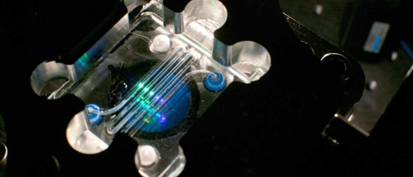
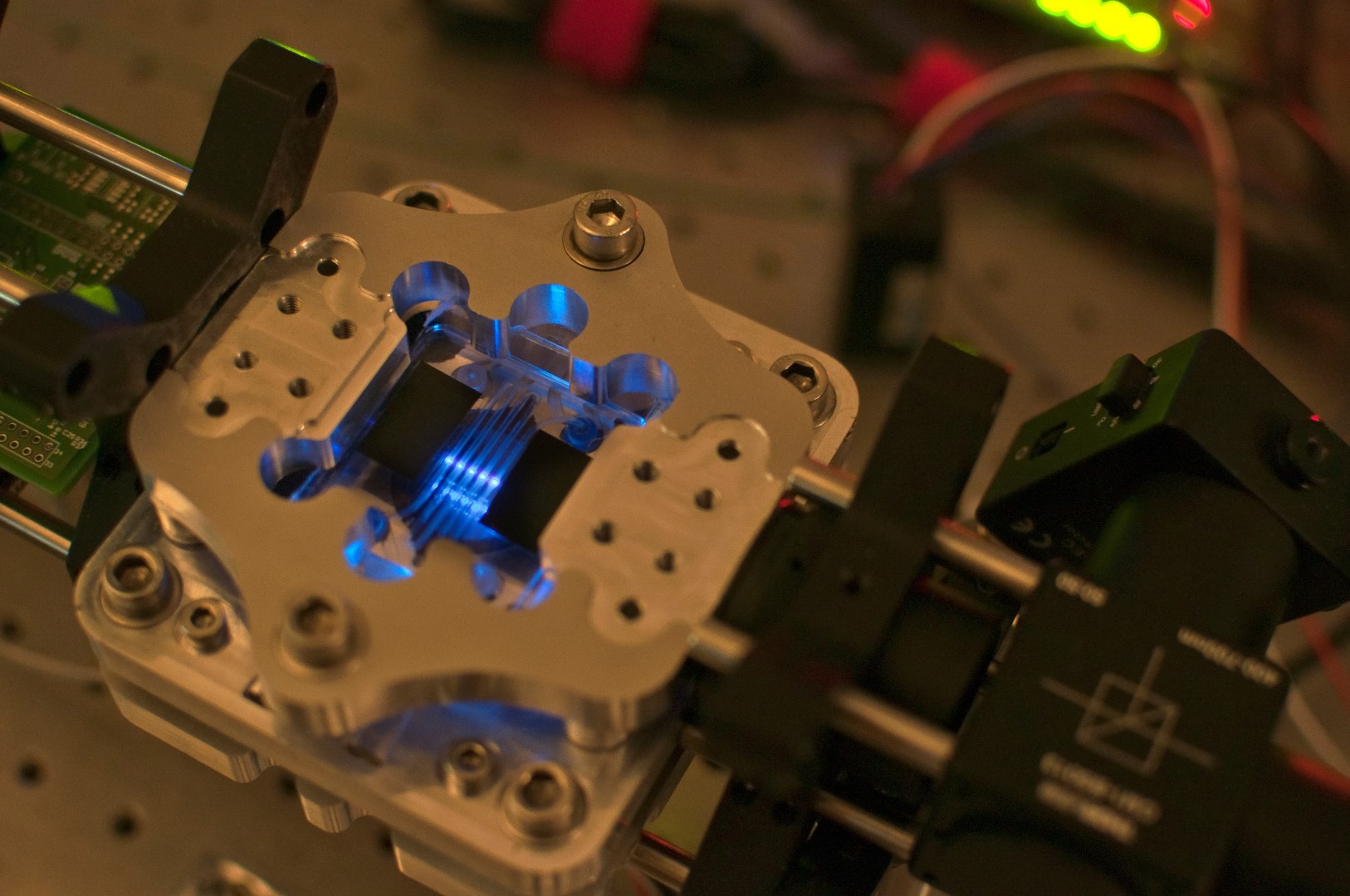
The B-PHOT photonics lab-on-chip team focuses on multifunctional opto-microfluidic systems that can combine optical absorption, fluorescence, surface-plasmon-resonance, and/or Raman spectroscopy in combination with optical trapping as on-chip optical detection techniques. These photonic labs-on-a-chip are fabricated in-house using B-PHOT’s highly advanced prototyping and pilot-line for polymer and glass micro-opto-fluidic components. Currently the team develops labs-on-chip for measuring the concentration of hazardous substances such as aflatoxins in food, the detection of pathogens in drinking water such as E-Coli and Salmonella, the authentication of olive oils, beer, honey or other products, and the testing of medical drugs using human-organs-on-chip to minimize the use of test-animals. Miniaturized optical spectroscopy systems are also designed for medical purposes ranging from the resistance/sensitivity screening of bacterial strains, the optical sensing of blood samples and human tissue for presence of free bilirubin, markers for Alzheimer’s disease, and carcinogenic cells, to the in-situ and real-time multimodal screening of organs-on-a-chip.
3D printing of microstructures for labs-on-chips
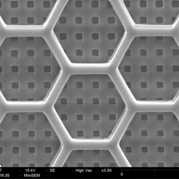
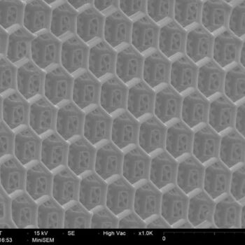
Our labs-on-chips can also be foreseen of scaffold structures to facilitate cell growth, which can then result in the realization of organs-on-chip. As an example, seeding and culturing hepatocyte-like cells in 3D scaffolds allow for the creation of an artificial liver on chip, which can then be used as an in-vitro testing platform for drug testing. By putting the cells in a 3D scaffold with microfluidic flow, one creates an environment wherein the cells are organised in a human-relevant configuration and will allow the model to be much more representative than cell cultures in Petri dishes without flow. In time, we want to contribute to reducing the need for using animals for drug testing.
Another important feature that can be added to microfluidic labs-on-chips by means of laser direct writing is nanostructured surfaces for surface-enhanced Raman spectroscopy (SERS). Since the intensity of Raman scattering is very low, working with SERS surfaces allows to strongly increase the Raman signal to allow a better detection.
Scaffolds
Scaffolds

Cell scaffolds
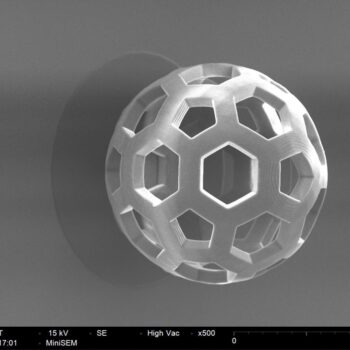
Scaffolds
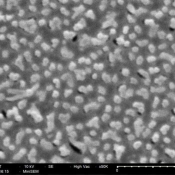
SERS surfaces
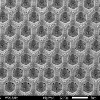
Chromatographic separation structures
Principal investigators
Post-docs
PhD Students


